Patellar Tendinosis
Anatomy
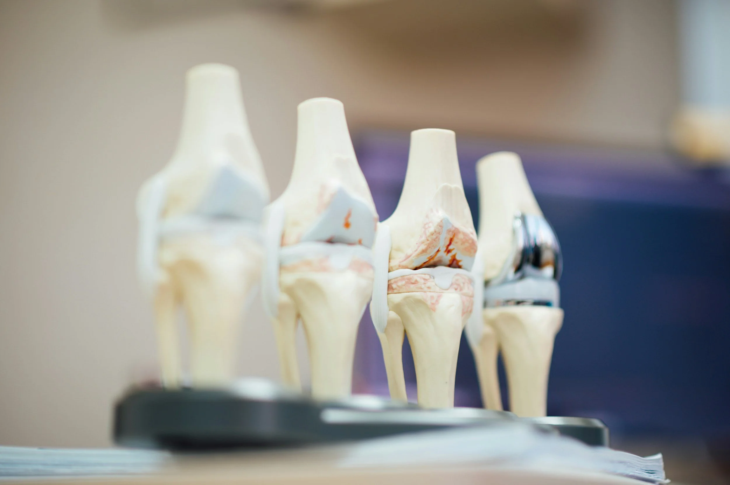
The knee functions as a hinge joint that allows bending and straightening of the leg. It consists of three bones: the femur (thigh bone), tibia (shin bone), and patella (kneecap).
The ends of these bones are covered with smooth articular cartilage, which reduces friction and helps absorb shock during movement. Between the femur and tibia are two structures called the medial and lateral menisci, which act as cushions and help distribute weight evenly across the joint.
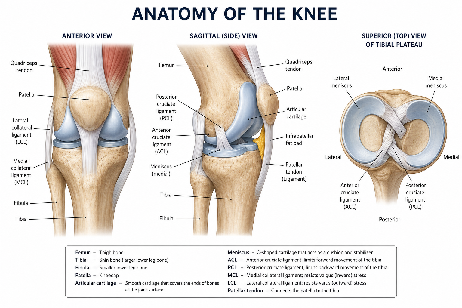
Stability of the knee is maintained by several important ligaments and surrounding muscles. The anterior cruciate ligament (ACL) and posterior cruciate ligament (PCL) control forward and backward movement of the tibia, while the medial collateral ligament (MCL) and lateral collateral ligament (LCL) stabilize the knee from side-to-side forces.
Muscles such as the quadriceps in the front of the thigh and the hamstrings in the back work together to move and support the joint. Tendons connect these muscles to the bones, allowing the knee to perform essential activities such as walking, running, jumping, and changing direction. The patellar tendon is a thick band of tissue that connects your kneecap to your shinbone and plays a key role in straightening the knee.
What is Patellar Tendinosis?
Patellar tendinosis is a chronic condition involving the breakdown of the patellar tendon.
Unlike a sudden injury, tendinosis develops from repeated stress on the tendon over time. This repetitive loading leads to a slow deterioration of the tendon's collagen fibers, leaving the tissue weakened, disorganized, and less able to handle the demands placed on it. It is especially common in athletes who frequently jump, sprint, or squat, which is why it is often nicknamed "jumper's knee."
The most common symptom is a dull, aching pain just below the kneecap that tends to worsen during or after physical activity. In the early stages, the pain may come and go, but without proper management, it can become persistent and significantly limit activity. As the tendon has a poor blood supply and thus heals slowly, recovery requires patience.
Treatment Options
Activity Modification: The first step in treatment is bringing the load on the tendon back to a manageable level by finding a training volume that the tendon can tolerate without worsening.
Physical Therapy: At times, broader mechanical issues are the reason for the tendon overload in the first place. A physical therapist can assess these factors and design a program that rehabilitates the tendon and corrects the movement patterns that contributed to the injury.
Platelet-Rich Plasma (PRP) Injection: PRP therapy involves injecting a concentration of the patient's own platelets into the diseased tendon to stimulate healing. It may be considered for chronic patellar tendinosis that has not improved with traditional conservative treatments.
Non-Operative Treatments
Surgical Options
Patellar Tendon Arthroscopic Debridement: The surgeon removes degenerative tendon tissue, scar tissue, and any areas of chronic inflammation within the patellar tendon. The goal is to promote healing of healthier tendon tissue and reduce pain.
Open Debridement with Tendon Repair: For more extensive tendon degeneration, an open procedure may be required. Diseased portions of the tendon are removed, and the remaining healthy tendon is repaired. In some cases, small drill holes are created in the patella to stimulate blood flow and healing.
Dr. Tingstad specializes in knee disorders and will help you select the best treatment based on your injury.