Patellar Instability
Anatomy
The knee functions as a hinge joint that allows bending and straightening of the leg. It consists of three bones: the femur (thigh bone), tibia (shin bone), and patella (kneecap).
The ends of these bones are covered with smooth articular cartilage, which reduces friction and helps absorb shock during movement. Between the femur and tibia are two structures called the medial and lateral menisci, which act as cushions and help distribute weight evenly across the joint.
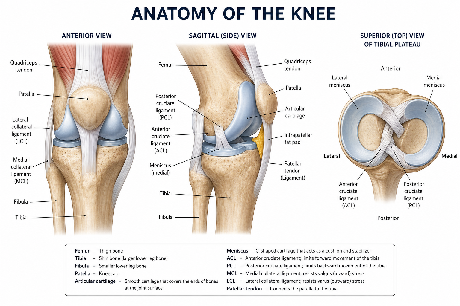
Stability of the knee is maintained by several important ligaments and surrounding muscles. The anterior cruciate ligament (ACL) and posterior cruciate ligament (PCL) control forward and backward movement of the tibia, while the medial collateral ligament (MCL) and lateral collateral ligament (LCL) stabilize the knee from side-to-side forces.
Muscles such as the quadriceps in the front of the thigh and the hamstrings in the back work together to move and support the joint. Tendons connect these muscles to the bones, allowing the knee to perform essential activities such as walking, running, jumping, and changing direction.
What is Patellar Instability?
Patellar instability occurs when the kneecap (patella) does not remain properly aligned within the groove at the end of the thigh bone (femur). This condition can cause the patella to partially slip out of place or completely dislocate, often resulting in pain, swelling, feelings of the knee "giving way," and difficulty with activity.
Instability can result from a traumatic injury such as a sudden twist or impact. It can also develop gradually due to underlying factors like a shallow groove, a high-riding kneecap, or weakness in the surrounding muscles. Once a dislocation has occurred, the supporting soft tissue structures are often damaged. This makes repeat episodes more likely, and over time, recurrent instability can lead to cartilage damage and early arthritis in the knee.
Treatment Options
Non-Operative Treatments
Strengthening exercises: One option to manage patellar instability without surgery is through a targeted exercise program. The goal is to strengthen the muscles around the knee to help keep the kneecap tracking in its groove. Strengthening the hip muscles is equally important, as weakness higher up the leg can place extra stress on the knee and contribute to the kneecap slipping out of place.
Bracing and taping: Wearing a knee brace can help prevent the kneecap from sliding sideways during everyday activities. A clinician may also apply a technique called McConnell taping, where firm tape is placed on the skin around the kneecap to hold it in a better position. Both approaches can ease pain and help patients exercise more effectively during the early stages of recovery.
Rest and gradual return to activity: In the short term, avoiding activities that put stress on the knee, such as pivoting, cutting movements, or deep squats, gives the injured soft tissues around the kneecap time to heal. Once symptoms settle, activity is slowly reintroduced in a structured way.
Surgical Options
MPFL reconstruction: The medial patellofemoral ligament (MPFL) is the primary soft tissue that stops the kneecap from sliding outward, and it is usually torn during a dislocation. Reconstruction involves using a tendon graft to recreate this ligament and restore stability.
Tibial tubercle osteotomy (TTO): In some patients, the bony attachment of the patellar tendon sits too far to the outside, pulling the kneecap laterally. A TTO involves cutting and repositioning this bony attachment to improve the alignment of the kneecap within its groove.
Trochleoplasty: The trochlea is the groove at the end of the thighbone that the kneecap sits in. In some patients this groove is abnormally shallow (trochlear dysplasia), meaning it provides little bony containment for the kneecap. Trochleoplasty is a procedure that deepens and reshapes this groove to improve stability.
Dr. Tingstad specializes in knee disorders and will help you select the best treatment based on your injury.