Knee Arthritis
Anatomy
The knee functions as a hinge joint that allows bending and straightening of the leg. It consists of three bones: the femur (thigh bone), tibia (shin bone), and patella (kneecap).
The ends of these bones are covered with smooth articular cartilage, which reduces friction and helps absorb shock during movement. Between the femur and tibia are two structures called the medial and lateral menisci, which act as cushions and help distribute weight evenly across the joint.
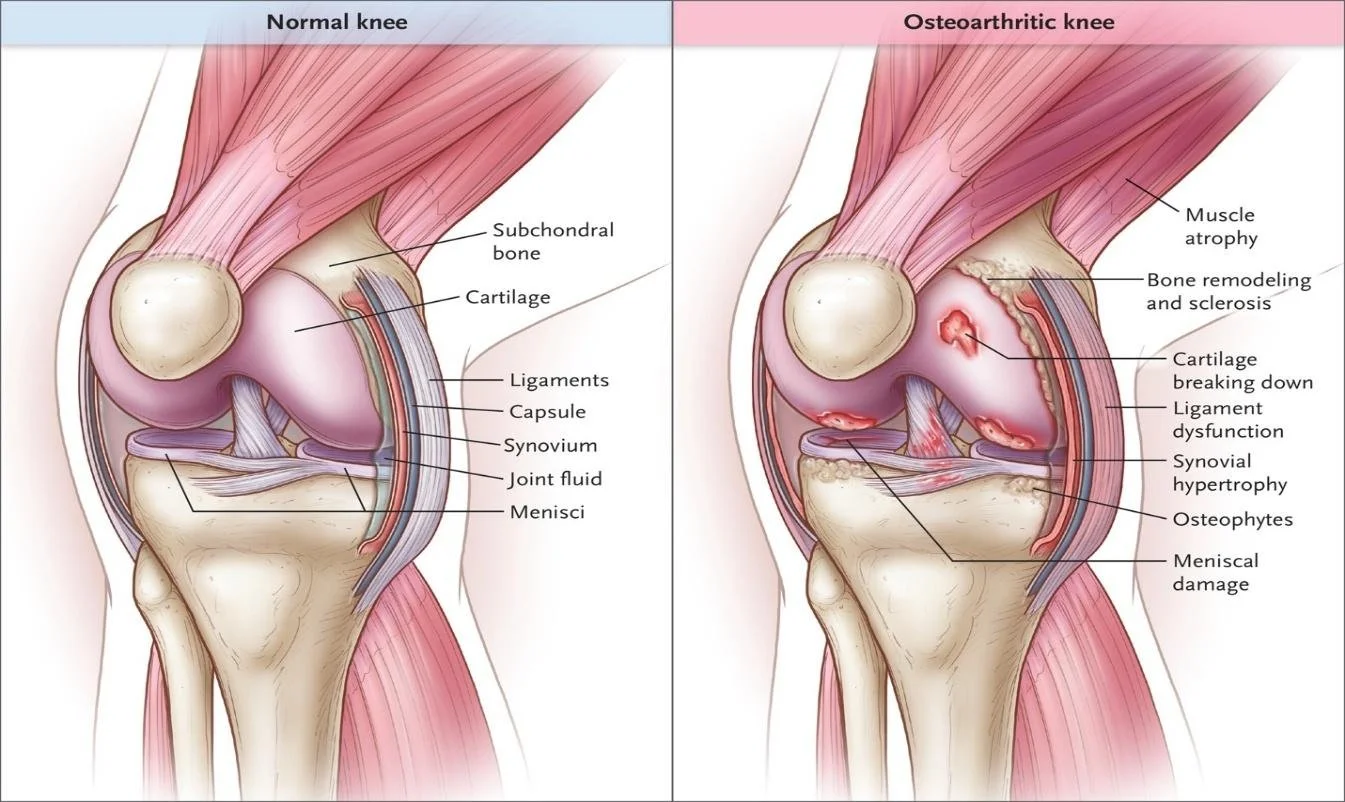
Stability of the knee is maintained by several important ligaments and surrounding muscles. The anterior cruciate ligament (ACL) and posterior cruciate ligament (PCL) control forward and backward movement of the tibia, while the medial collateral ligament (MCL) and lateral collateral ligament (LCL) stabilize the knee from side-to-side forces.
Muscles such as the quadriceps in the front of the thigh and the hamstrings in the back work together to move and support the joint. Tendons connect these muscles to the bones, allowing the knee to perform essential activities such as walking, running, jumping, and changing direction.
What is Knee Arthritis?
Knee arthritis happens when the cartilage cushioning the bones in your knee joint wears down over time. That cartilage normally lets the bones glide against each other smoothly. Once it thins, the bones move closer together, and that closeness is what causes the stiffness, swelling, and pain tied to arthritis. Osteoarthritis is the most common form.
Osteoarthritis tends to develop slowly with age, though injuries, extra weight on the joints, and family history can accelerate this timing.
Symptoms tend to build slowly, often worsening with activity like standing, walking, or climbing stairs. Many people feel stiff right after waking or after sitting for a long stretch, and a grinding or clicking sensation can show up when the knee bends or straightens. Swelling around the joint is common.
Treatment Options
Non-Operative Options
Corticosteroid injections provide temporary pain relief by reducing inflammation and improving the body’s ability to manage the inflammation in the joint.
Visco supplementation injections aim to relieve pain by lubricating the joint with a gel-like fluid. This fluid allows the articulating surfaces of the joint to move more smoothly causing less inflammation.
Physical therapy is recommended to increase your range of motion and coordination of muscles to better support the knee.
NSAID/anti-inflammatory medications can provide pain relief and decrease inflammation around the knee.
Medial/lateral unloader braces offload areas of osteoarthritis and prevent inwards collapse of the knee by applying pressure and aligning the knee into a neutral position.
Surgical Options
Arthroscopy is a minimally invasive procedure to clean out loose cartilage or debris.
Osteotomy, where a surgeon reshapes the bone to shift weight away from the damaged part of the knee, is often considered for younger or more active patients with arthritis isolated to one side of the joint.
Partial (unicompartmental) knee replacement, replaces just the damaged section of the knee.
Total knee replacement, replacing the entire joint surface, which is the most common surgical option for advanced arthritis.
Cartilage restoration procedures like microfracture or osteochondral grafting are generally reserved for younger patients with smaller, more localized cartilage damage rather than widespread arthritis.
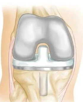
Shown above: a total knee replacement
Dr. Tingstad specializes in treating knee arthritis and will help you select the best treatment based on your injury.