Elbow Arthroscopy
Anatomy
The elbow is a joint formed by three bones: the humerus in the upper arm, and the radius and ulna in the forearm. It allows two types of movement: bending and straightening the arm, and rotating the forearm to turn the palm up or down. The joint is stabilized by ligaments on both sides. On the inner side is the ulnar collateral ligament (UCL). On the outer side is the lateral collateral ligament (LCL).
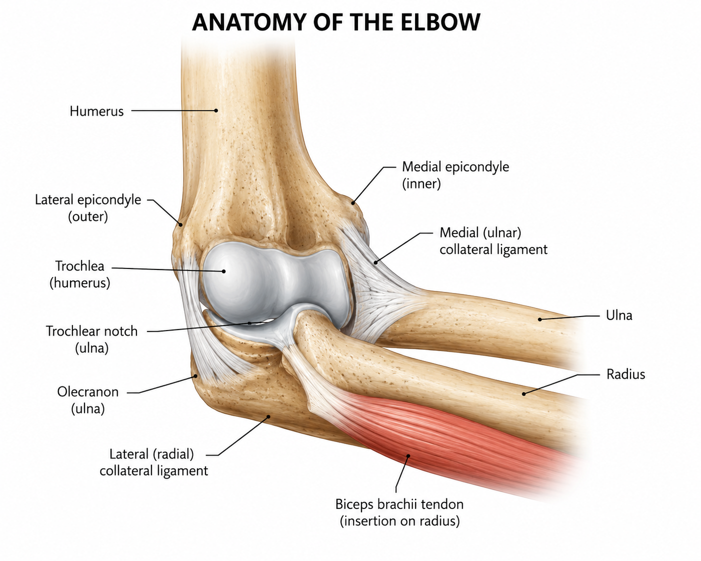
Several muscle groups cross the elbow. The biceps and brachialis bend the elbow, and the triceps straighten it. The forearm muscles that move the wrist and fingers also attach at the elbow, at bony points called the medial and lateral epicondyles. The medial epicondyle is on the inner side, where the ulnar nerve passes close to the surface. The lateral epicondyle is on the outer side, where the wrist extensor muscles attach.
Arthroscopy
Elbow arthroscopy is a minimally invasive surgical procedure used to diagnose and treat a variety of conditions affecting the elbow joint. During the procedure, a surgeon makes several small incisions around the elbow and inserts a specialized camera called an arthroscope into the joint.
The arthroscope provides a magnified view of the elbow's internal structures, allowing the surgeon to identify and address problems without the need for a large open incision. Specialized instruments can then be introduced through additional small portals to perform necessary treatments.
Elbow arthroscopy can be used to treat numerous conditions, including loose bodies, elbow stiffness, cartilage injuries, arthritis, and certain sports-related injuries. Compared to traditional open surgery, arthroscopy often results in less postoperative pain, smaller scars, reduced soft tissue disruption, and a faster recovery.