Articular Cartilage Defects and/or Osteochondral Defects
Anatomy
The knee functions as a hinge joint that allows bending and straightening of the leg. It consists of three bones: the femur (thigh bone), tibia (shin bone), and patella (kneecap).
The ends of these bones are covered with smooth articular cartilage, which reduces friction and helps absorb shock during movement. Between the femur and tibia are two structures called the medial and lateral menisci, which act as cushions and help distribute weight evenly across the joint.
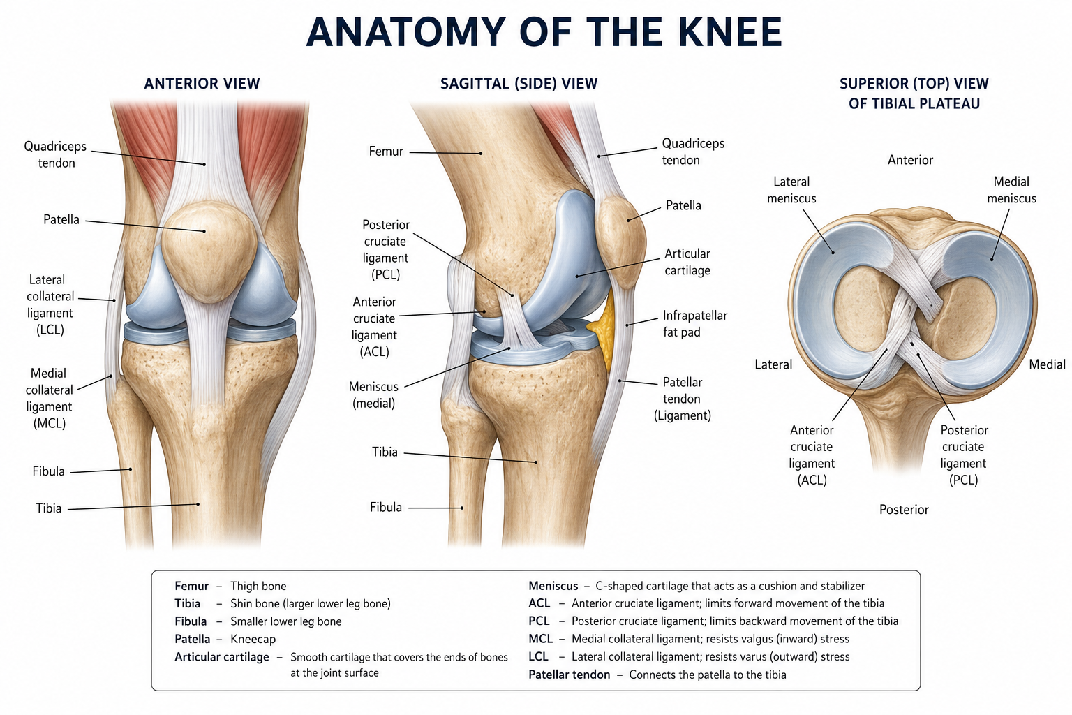
Stability of the knee is maintained by several important ligaments and surrounding muscles. The anterior cruciate ligament (ACL) and posterior cruciate ligament (PCL) control forward and backward movement of the tibia, while the medial collateral ligament (MCL) and lateral collateral ligament (LCL) stabilize the knee from side-to-side forces.
Muscles such as the quadriceps in the front of the thigh and the hamstrings in the back work together to move and support the joint. Tendons connect these muscles to the bones, allowing the knee to perform essential activities such as walking, running, jumping, and changing direction.
What are Articular Cartilage Defects?
Articular cartilage is the smooth, white tissue that covers the ends of bones within joints, allowing them to glide against one another with minimal friction. Articular cartilage defects are localized areas of damage or loss confined to this cartilage layer, without extending into the underlying bone.
These defects typically arise from acute trauma, repetitive mechanical stress, or degenerative conditions, and they are particularly common in the knee. Articular cartilage lacks a direct blood supply and therefore has a very limited capacity for self-repair. Even small defects can worsen over time, causing pain, swelling, and mechanical dysfunction, and if left untreated, they can eventually contribute to the development of osteoarthritis.
What are Osteochondral Defects?
Osteochondral defects are a more severe form of joint injury in which damage extends beyond the articular cartilage and into the subchondral bone beneath it. These defects can result from trauma, osteochondritis dissecans (a condition where a fragment of bone and cartilage separates from the joint surface), or progressive degeneration. Osteochondral defects are generally more complex to treat than purely cartilage defects and often require surgical intervention.
Treatment Options
Non-Operative Treatment of Articular Cartilage Defects
Physical therapy to strengthen the muscles around the affected joint and reduce stress on the damaged cartilage
Activity modification, such as avoiding high-impact activities like running or jumping, to prevent further deterioration
NSAIDs or corticosteroid injections to manage pain and inflammation
Viscosupplementation, which involves injecting hyaluronic acid into the joint to improve lubrication
Bracing to offload the affected area of the joint and provide stability
Surgical Options for Articular Cartilage Defects
Microfracture: A technique where small holes are made in the bone beneath the defect to stimulate the release of bone marrow cells and encourage the formation of repair tissue.
Matrix Autologous Chondrocyte Implantation (MACI): A two-stage procedure where cartilage cells are harvested from the patient, grown in a lab, and then implanted into the defect.
Osteochondral Autograft Transfer (OATS): Healthy cartilage and bone plugs are taken from a non-load-bearing area of the joint and transplanted into the defect.
Particulated juvenile cartilage allograft(DeNovo NT): This procedure uses donor cartilage tissue to fill the defect and promote healing.
Scaffold-based techniques: This procedure uses biocompatible materials to support the growth of new cartilage tissue within the defect.
Non-Operative Treatment of Osteochondral Defects
Protected weight bearing or rest, particularly in younger patients, to allow for any natural healing response in the bone layer.
Physical therapy to maintain joint function and muscle strength during recovery.
NSAIDs or corticosteroid injections to control pain and swelling.
Bracing or orthotics to redistribute the load across the joint.
Platelet-rich plasma (PRP) injections are an emerging option to encourage tissue healing.
Surgical Options for Osteochondral Defects
Microfracture: A technique where small holes are made in the bone beneath the defect to stimulate the release of bone marrow cells and encourage the formation of repair tissue.
Osteochondral Autograft Transfer (OATS): Plugs of healthy cartilage and bone are taken from the patient and used to fill the defect.
Osteochondral Allograft Transplantation: The surgeon uses donor bone and cartilage to replace the damaged tissue.
Bone grafting procedures: The procedure addresses the bone-loss component of the defect before or alongside cartilage restoration.
Matrix Autologous Chondrocyte Implantation (MACI): This may be used in combination with bone grafting for defects that involve both cartilage and bone.
Dr. Tingstad specializes in meniscus injuries and will help you select the best treatment based on your injury.