Anterior Cruciate Ligament (ACL)
Anatomy
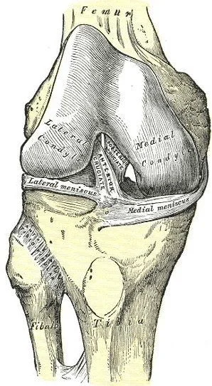
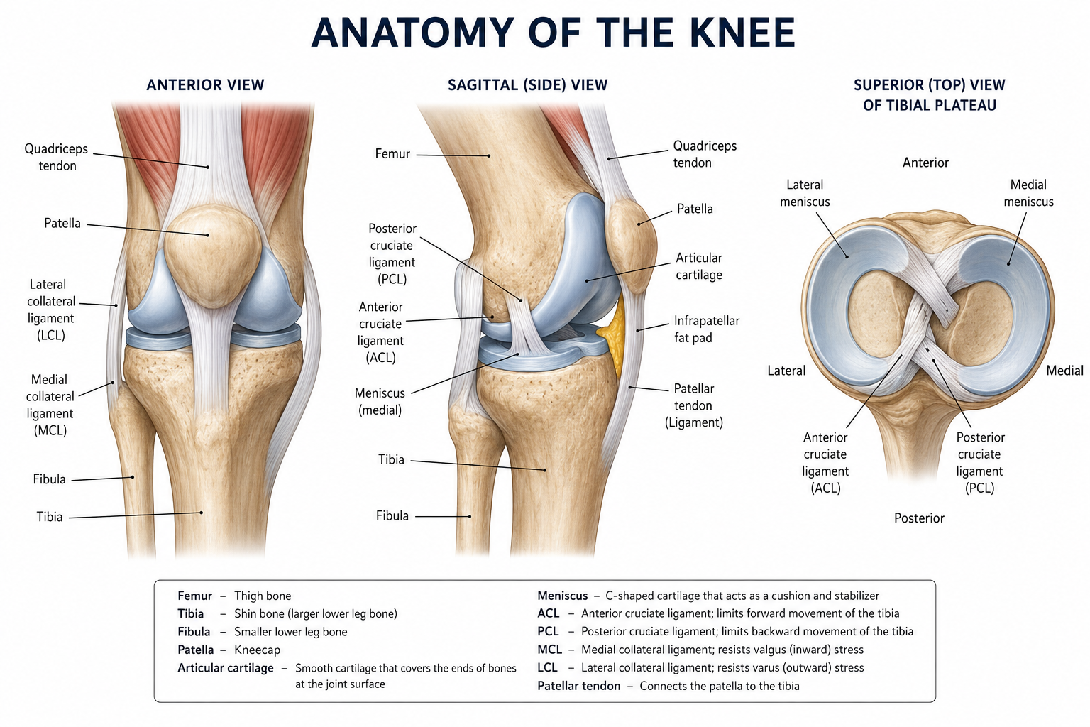
Four major ligaments provide stability to the knee. They are the anterior cruciate ligament (ACL), the posterior cruciate ligament (PCL), the medial collateral ligament (MCL), and the lateral collateral ligament (LCL).
The ACL is located inside the knee joint next to the PCL. The MCL and LCL are on the outside of the knee joint, and the PCL is inside the knee near the back. There are also two meniscal cartilages that act as shock absorbers and provide some stability. The ACL lines the knee joint and allows for smooth, nearly frictionless motion.
A torn ACL is commonly associated with injury to two or more of these structures.
A Torn ACL
Injuries to the anterior cruciate ligament (ACL) occur most often in athletic activities. The injury typically does not require a blow to the knee but instead involves a rapid change in direction or a deceleration maneuver. Almost 70% of ACL injuries are not associated with any contact.
The individual will often hear or feel a “pop” inside the knee. The injury is usually followed by increasing pain and swelling, as well as progressive inability to bear weight on the leg, for a short period of time. However, some people will swell very little after an ACL injury.
Treatment Options
The decision to undergo reconstructive surgery for an ACL tear is highly individualized. Patients who plan to continue an active lifestyle should consider undergoing ACL reconstruction.
Reconstruction of the ACL is a surgical procedure performed in an operating room, usually under general or spinal anesthesia. The ligament reconstruction is performed by removing the remnant of the torn ACL from the knee, then replacing it with biologic tissue.
The replacement may come from one of several sources (see photo on the right). These include your patellar tendon or hamstring tendons (called “autografts”) or tissue from tissue banks (called “allografts”). Alternatively, certain patients may choose to undergo the BEAR ACL procedure instead of a graft reconstruction.
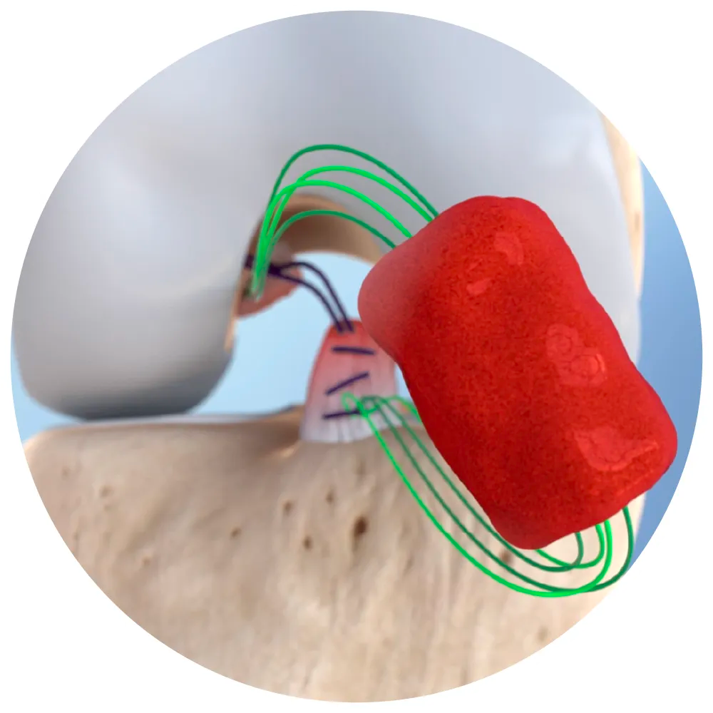
During the BEAR procedure, the surgeon sutures a bovine-collagen implant between the torn ends of the ACL. The implant acts as a “super clot” to help the ACL repair itself naturally. This option is less invasive and preserves natural knee joint function.
Dr. Tingstad specializes in all types of ACL procedures and will discuss options with you during your visit.
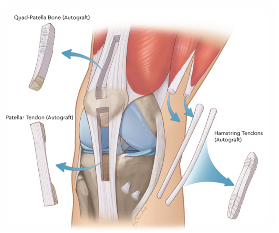
Above: ACL Reconstruction Graft Options
Below: BEAR ACL implant with sutures on both ends of torn ACL