Achilles Tendon Injuries
Anatomy
The Achilles tendon is the largest and strongest tendon in the body, connecting the calf muscles, the gastrocnemius and soleus, to the heel bone (calcaneus). This powerful tendon plays a critical role in walking, running, jumping, and pushing off the foot during movement.
During athletic activities, the Achilles tendon is subjected to significant forces, making it vulnerable to conditions such as tendinitis, tendinosis, and tendon rupture. Its relatively limited blood supply in certain regions can also contribute to slower healing following injury.
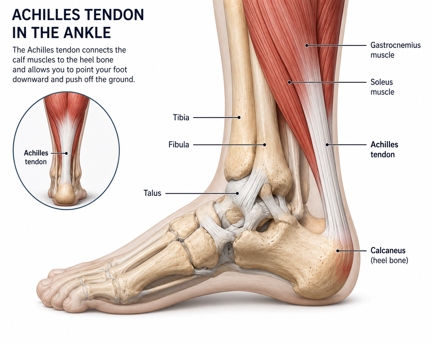
The ankle is a complex joint composed primarily of the tibia, fibula, and talus, which work together to provide stability while allowing motion in multiple directions. Numerous ligaments support the ankle, helping maintain alignment and prevent excessive movement, while surrounding muscles and tendons contribute to strength and balance.
The ankle and Achilles tendon function as an integrated system, enabling efficient movement, shock absorption, and propulsion during everyday activities and sports. Proper coordination among these structures is essential for maintaining mobility, stability, and overall lower extremity function.
Achilles Tendon Injuries
Achilles Tendinopathy: This refers to a gradual breakdown of the tendon tissue due to overuse and repetitive loading. It typically causes pain and stiffness at the back of the heel, which is often worse first thing in the morning or at the start of exercise and eases as the tendon warms up.
Tendinopathy is particularly common in runners and other athletes who have recently increased their training load.
Achilles Tendon Rupture: A rupture is a partial or complete tear of the Achilles tendon, most often occurring in the vulnerable zone just above the heel. It typically happens during a sudden explosive movement such as sprinting, jumping, or pushing off forcefully. Patients often describe hearing or feeling a loud "pop," as if they had been kicked in the back of the leg.
Treatment Options
Non-Operative Options
Eccentric Exercise (Heel-Drop Program): A structured 12-week exercise program focused on slowly lowering the heel off a step is the most effective first-line treatment. It works by stimulating the tendon to rebuild healthier tissue over time.
Physical Therapy: A physical therapist addresses strength and movement issues in the calf, hip, and foot that may be contributing to the problem, reducing symptoms and preventing recurrence.
Shockwave Therapy (ESWT): A device delivers pressure waves to the tendon to promote healing. This is particularly useful for stubborn cases that haven't responded to exercise alone.
Heel Lifts & Orthotics: Simple shoe inserts or heel lifts reduce the load on the tendon during daily activity and can help ease symptoms early on.
NSAIDs: Over-the-counter anti-inflammatories like ibuprofen can reduce short-term pain.
Functional Bracing: A walking boot with a raised heel is used to hold the tendon ends close together while the tendon heals naturally.
Surgical Options
Open Surgical Repair: The two ends of the torn tendon are sutured back together through an incision at the back of the ankle. This is the traditional approach and offers a low re-rupture rate.
Minimally Invasive Repair: The tendon is repaired through small incisions rather than one large opening, reducing the risk of wound complications and nerve injury while still directly reconnecting the tendon.
Reconstruction (Chronic/Neglected Tears): If the rupture is diagnosed late and the tendon ends have retracted too far to be sutured together, more complex surgery is needed. The surgeon often uses the patient's own tissue to bridge the gap and rebuild the tendon.